IVDD Recovery Timeline: From Diagnosis to Walking Again
IVDD Recovery in Dogs:
From Diagnosis to Walking Again
Author: Karen Goodall AdvCertVPhys MIRVAP (VP) (ICH) | Veterinary Physiotherapist, Old Flatts Farm, Treeton
What Is IVDD — and Why Do Timelines Vary?
Intervertebral Disc Disease (IVDD) occurs when the cushioning disc between two vertebrae either bulges gradually (Hansen Type II) or ruptures suddenly, sending disc material into the spinal canal (Hansen Type I). This compresses the spinal cord and nerve roots, causing a spectrum of clinical signs ranging from neck or back pain to hindlimb weakness, incoordination (ataxia), and in severe cases, paralysis and loss of bladder or bowel control.
Certain breeds are disproportionately affected due to a genetic process called chondrodystrophy, which causes premature disc degeneration. Dachshunds account for approximately 45–70% of all IVDD cases (Smolders et al., 2013), with French Bulldogs, Corgis, Beagles, and Cocker Spaniels also at elevated risk. Large and giant breeds can also be affected, typically presenting with slower-onset disc protrusion rather than acute extrusion.
Advanced imaging — MRI or CT — is required to confirm diagnosis, locate the compression, and guide treatment decisions. Neurological grade at the time of presentation is one of the strongest predictors of outcome (Olby et al., 2003).
Treatment Paths
- Surgical decompression (e.g., hemilaminectomy, ventral slot) is generally recommended for dogs with moderate to severe neurological deficits, rapidly deteriorating function, or failure to respond to conservative management. Surgery removes the offending disc material to relieve spinal cord compression.
- Conservative management involves strict activity restriction, anti-inflammatory and analgesic medication, bladder management if needed, and a carefully staged rehabilitation programme. It is most appropriate for dogs with mild to moderate neurological signs, or where surgery is not possible.
Because the two pathways follow different pacing — and because neurological grade, lesion location, and individual response all vary — the timelines below are guides rather than guarantees. Your veterinary team is best placed to predict your dog’s individual outlook.
Phase 1: Stabilise and Protect (Weeks 0–2)
What This Looks Like at Home
- Strict rest is essential — think of it as the invisible cast. Your dog’s spine needs the same protection a broken bone would receive
- Short, controlled lead walks for toileting only — support with a well-fitted harness or sling if your dog is wobbly or weak
- Non-slip flooring throughout the home; remove access to stairs, sofas, and beds
- Administer prescribed medication on schedule — pain management and anti-inflammatories are critical in this phase
- Monitor bladder function closely; some dogs lose voluntary bladder control and may require assisted expression
- For post-operative dogs: follow incision care instructions carefully and contact your vet with any wound concerns
Physiotherapy Focus
Where appropriate and under veterinary direction, a physiotherapist may introduce gentle passive range of motion (PROM) to the limbs to maintain joint mobility and reduce muscle stiffness. Soft tissue massage can help relieve the significant paraspinal muscle guarding that accompanies spinal pain. In post-operative cases, assisted positioning, handling, and supported standing may begin during this phase.
Owner education is a significant component of this phase — correct handling, sling use, assisted toileting, and knowing what to monitor at home all reduce the risk of deterioration and improve outcomes.
Phase 2: Early Rehabilitation (Weeks 2–4)
What This Looks Like at Home
- Continue strict activity restriction as directed — many dogs remain on crate rest at this stage
- Begin prescribed home exercises: these may include assisted standing, gentle limb cycling, or supported balance work depending on neurological grade
- Keep a simple rehabilitation diary — note exercise sessions, repetitions, any signs of fatigue or discomfort, and changes in gait
- Maintain non-slip surfaces for all exercise and toileting
Physiotherapy Focus
This phase introduces more active rehabilitation. The physiotherapist will assess neurological status, postural reactions, and movement quality to guide progression. Techniques commonly used include neuromuscular electrical stimulation (NMES) to support activation of weak or paretic limbs, therapeutic laser (photobiomodulation) for tissue healing and pain modulation, targeted range of motion and joint mobilisation, and postural response facilitation exercises.
Evidence for individual techniques used in isolation remains limited; however, current reviews support a multimodal, patient-specific rehabilitation approach overseen by a qualified professional as the most effective strategy (Levine et al., 2007; Sharp and Wheeler, 2005). The physiotherapist will select and combine modalities based on your dog’s presentation and response.
Hydrotherapy or underwater treadmill therapy may be introduced at this stage for suitable cases — the buoyancy of water reduces spinal load while enabling active limb movement, which is particularly valuable for dogs with hindlimb weakness. Surgeon clearance and appropriate wound healing are required before hydrotherapy begins.
Phase 3: Strength, Balance and Gait (Weeks 4–8)
What This Looks Like at Home
- Gradually increasing, controlled lead walks as directed — short and frequent is preferable to long and infrequent
- Home exercise programme progresses to include balance challenges, sit-to-stand repetitions, and slow stepping over cavaletti poles
- Continue to avoid jumping, stairs, and off-lead activity until cleared by your veterinary team
- Monitor carefully: no limping during or after exercise, no increased stiffness the following morning
- Weight management is important at this stage — excess bodyweight significantly increases spinal loading (Packer et al., 2013)
Physiotherapy Focus
Proprioception — the nervous system’s ability to detect and respond to limb position — is frequently disrupted following spinal cord injury. Targeted exercises on varied surfaces, balance boards, and dynamic standing tasks help re-establish these pathways. Recovery of conscious proprioception is a positive prognostic indicator and an important marker of rehabilitation progress (Olby et al., 2003).
The physiotherapist will use objective measures — observational gait analysis, postural reaction testing, and muscle girth measurements — to track progress and identify any compensatory patterns developing in the forelimbs, neck, or contralateral hindlimb. Spinal cord injuries place significant additional demand on the thoracic limbs, and targeted soft tissue work and strengthening for these regions is an important but often overlooked component of spinal rehabilitation.
Phase 4: Return to Function (Weeks 8–16+)
What This Looks Like at Home
- Gradual return to normal walks and home routine — off-lead exercise is reintroduced cautiously and only when cleared
- Permanent environmental modifications are strongly recommended: ramps for furniture and car access, stair gates, non-slip flooring throughout
- Use a harness for all walks, even after full recovery — collars place unnecessary load on the cervical spine
- Maintain appropriate exercise and weight management long-term
- For Dachshunds and other high-risk breeds: consider a spinal health check as part of annual veterinary care
Physiotherapy Focus
The focus shifts to maintenance and recurrence prevention. A structured home exercise programme of 2–3 sessions per week helps sustain neuromuscular gains. Periodic physiotherapy check-ins allow for programme refinement as your dog’s activity level increases.
Recurrence rates for IVDD are significant — studies suggest that approximately 30–40% of dogs managed conservatively experience a recurrence within two years (Smolders et al., 2013). Long-term physiotherapy support, alongside weight management, appropriate exercise, and environmental adaptations, forms the foundation of a recurrence-prevention strategy.
For non-ambulatory dogs, recovery can be a longer journey. Many dogs — even those initially unable to walk — regain function with dedicated rehabilitation, committed owners, and time. Deep pain perception at the time of presentation remains the most reliable indicator of long-term prognosis (Olby et al., 2003), and your vet team will guide realistic goal-setting based on this finding.
Crate Rest: The Invisible Cast
Strict activity restriction — typically 4–6 weeks for conservative cases and 6–8 weeks post-operatively — is one of the most important, and most challenging, aspects of IVDD management. Think of it as the equivalent of a plaster cast for a broken bone: healing tissues cannot recover effectively if they are continually disturbed.
Practical Tips for Crate Rest
- Choose a crate or pen large enough for your dog to stand, turn around, and lie fully stretched — but not so large that they can build up pace
- Line with non-slip bedding; orthopaedic memory foam is ideal for spinal cases
- Raise food and water bowls, especially for cervical (neck) IVDD, to reduce spinal flexion during feeding
- Provide calm mental enrichment: lick mats, sniff work from a stationary position, and calm companionship
- Carry your dog outside for toileting rather than allowing unsupported walking where possible
- Always use a well-fitted, supportive harness for any assisted movement — never a collar
Owner compliance with rest significantly influences outcome. It is one of the hardest parts of IVDD management for many families, but also one of the most impactful. Your physiotherapist and vet team are there to support you through this stage.
Conservative vs Surgical Recovery: What to Expect
The two treatment pathways share the same rehabilitation principles but differ in pacing and prognosis. The table below outlines the key differences to help manage expectations:
| Conservative Management | Post-Surgical Recovery |
|---|---|
|
|
Frequently Asked Questions
How soon should physiotherapy start?
Ideally within the first 1–2 weeks of diagnosis or surgery, under veterinary direction. Early rehabilitation — even gentle positioning, passive movement, and sensory stimulation — has been shown to support neurological recovery and reduce muscle loss. A pre-operative assessment, where possible, provides useful baseline data to inform the rehabilitation programme.
Is hydrotherapy always appropriate for IVDD?
Hydrotherapy can be an excellent adjunct for spinal rehabilitation — the buoyancy of water reduces loading on the spine while enabling active limb movement and building confidence in dogs with hindlimb weakness. However, it is not suitable for every dog at every stage. Wound status, neurological grade, individual tolerance, and case selection all influence timing and suitability. Your physiotherapist and vet will guide this decision together.
My dog still has weakness at 8 weeks — should I be worried?
Not necessarily. Neurological recovery can be slow and non-linear — dogs commonly continue improving for many months after injury or surgery. Those with more severe spinal cord involvement naturally take longer to recover. The most important indicator is continued, steady progress. If you notice a plateau or regression, discuss this with your vet and physiotherapist so the programme can be reviewed.
What are the red flags during rehabilitation?
- Sudden or rapid worsening of neurological signs
- New loss of deep pain sensation in the hindlimbs
- Acute inability to urinate or unexpected loss of bladder or bowel control
- Wound swelling, discharge, or breakdown in post-operative cases
- Behavioural signs of significant pain: vocalisation, changes in appetite, or reluctance to move
Any of the above warrant immediate contact with your veterinary surgeon.
Can IVDD recur?
Yes — recurrence is a genuine risk, particularly in chondrodystrophic breeds. This is why long-term physiotherapy support, weight management, environmental adaptations, and appropriate exercise are not optional extras but core components of ongoing spinal health management. Your physiotherapist can discuss specific strategies to reduce recurrence risk based on your dog’s breed, injury severity, and lifestyle.
Scientific References
Levine, J.M., Levine, G.J., Johnson, S.I., Kerwin, S.C., Hettlich, B.F. and Fosgate, G.T. (2007). Evaluation of the success of medical management for presumptive thoracolumbar intervertebral disk herniation in dogs. Veterinary Surgery, 36(5), 482–491.
Levine, D., Marcellin-Little, D.J., Millis, D.L., Tragauer, V. and Osborne, J.A. (2008). Effects of partial immersion in water on vertical ground reaction forces and weight distribution in dogs. American Journal of Veterinary Research, 69(10), 1357–1363.
Olby, N., Levine, J., Harris, T., Munana, K., Skeen, T. and Sharp, N. (2003). Long-term functional outcome of dogs with severe injuries of the thoracolumbar spinal cord: 87 cases (1996–2001). Journal of the American Veterinary Medical Association, 222(6), 762–769.
Packer, R.M.A., Hendricks, A., Volk, H.A., Shihab, N.K. and Burn, C.C. (2013). How long and low can you go? Effect of conformation on the risk of thoracolumbar intervertebral disc extrusion in domestic dogs. PLOS ONE, 8(7), e69650.
Sharp, N.J.H. and Wheeler, S.J. (2005). Small Animal Spinal Disorders: Diagnosis and Surgery (2nd ed.). Elsevier Mosby.
Smolders, L.A., Bergknut, N., Grinwis, G.C.M., Hagman, R., Lagerstedt, A.S., Hazewinkel, H.A.W., Tryfonidou, M.A. and Meij, B.P. (2013). Intervertebral disc degeneration in the dog. Part 2: Chondrodystrophic and non-chondrodystrophic breeds. Veterinary Journal, 195(3), 292–299.
Book a Spinal Rehabilitation Assessment
If your dog has been diagnosed with IVDD — or is recovering after spinal surgery — we will design a tailored rehabilitation plan that fits your dog’s neurological grade, surgical outcome, and home routine. Our clinic is based at Old Flatts Farm, Treeton (Rotherham/Sheffield).
Request an Appointment →IVDD recovery guide
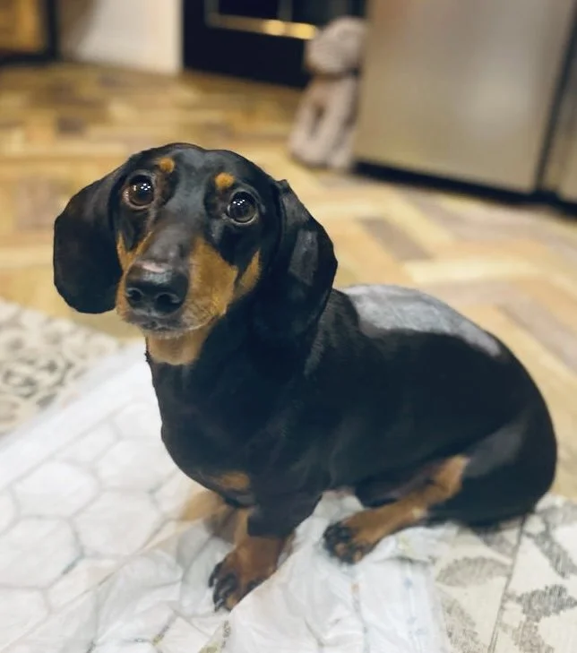
Post-operative canine IVDD patient